Our services
MRI
The development of MRI has revolutionised the medical world. Since its discovery, doctors and researchers have refined techniques to use MRI scans to assist in medical procedures and also help in research. In particular, MRI can be very important in the detection of prostate cancer.
What is an MRI?
An MRI scan does not use x-rays. Instead, it uses magnetic fields and radio waves to create a detailed image of the body’s tissues and structures. Doctors often use it to provide additional information to other tests, such as x-rays or ultrasound.
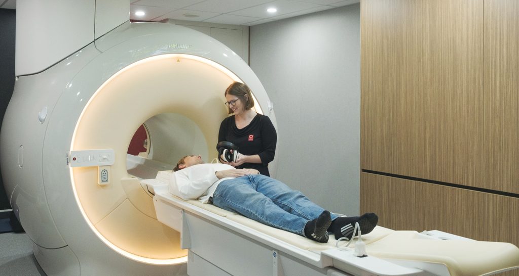
As you lay on the bed, you will feel it moving smoothly along the tracks, gliding into the center of the tunnel. Cameras line the walls around you, ready to capture the perfect shot of you in this unique setting.
Doctors can carry out many different types of MRI scans. Each one offers our team of Radiologists specific information to help diagnose the medical condition you might have.

Why is an MRI done?
MRI is used to capture images of a variety of body parts and bodily functions to diagnose a range of issues.
MRI of the breast offers valuable information about many breast conditions that cannot be obtained by other imaging, such as mammography or ultrasound. Breast MRI does not use ionizing radiation and provides a method for imaging related to silicone breast implants.
What are the advantages of Breast MRI?
MRI is the most sensitive method for diagnosing breast cancer and may be an appropriate screening tool for young women who have a high risk of breast cancer. Young women tend to have dense breast tissue, for which MRI is more appropriate than x-ray mammography. Because of its high accuracy in tissue differentiation, MRI is also becoming more and more important for staging breast cancer, which is crucial to determining the most appropriate treatment.
Is Breast MRI right for you?
Close consultation with your GP and Specialist will assess the need for you to have MRI of your Breasts. Because MRI does not use ionising radiation and is very sensitive to soft tissue change it can be a useful tool in diagnosing and staging breast disease and in tailoring treatment plans.
The prostate is a small organ located deep in the pelvis that has been traditionally difficult to image successfully in the detection of prostatic cancer. However, the use of next generation 3T MRI scanners and special parameters has made the detection of prostatic cancers much more accurate. Sunshine Coast Radiology is proud to offer the Sunshine Coast the only private next generation Philip’s Multi-parametric 3T MRI scanner. All exams are designed and interpreted by our team of RANZCR trained Radiologists who possess extensive experience in Prostate MRI scanning.
Is Multi-Parametric 3T MRI right for you?
Close consultation with your GP and Specialist will assess the need for you to have a 3T mulit-parametric MRI scan of your prostate. Certain high-risk individuals for prostate cancer could benefit by Multi-parametric 3T MRI. It can be useful for pre-operative staging and treatment plans and useful post-prostatectomy, radiation, or other treatment, in detecting recurrent disease.

About Your Test
Before your MRI appointment
You will need a referral from your doctor to make an appointment.
Upon receiving your referral, our Bookings Team will be able to help assist you in finding a time that works for you to have your test done.
Prior to the MRI scan you will be required to complete a safety questionnaire. Any preparation required will be communicated when you make your booking.
If you are pregnant or have a pacemaker or other implant it is important to tell the Booking Team when making your appointment.
If you a claustrophobic, advise your referring doctor and our team when making your appointment. Your doctor may prescribe a sedative medication.
On the day
On the day of your appointment you may be required to fast or undertake a special preparatory diet.
It is recommended you do not wear any makeup or hairspray as these products can contain small metal particle that can interfere with the quality of the MRI images.
You will be asked to remove all jewellery and any clothing items with metal parts (zips, eyelets, clips, underwires and hooks).
The length of the MRI will depend on the procedure but typically lasts 15-60 minutes. Your radiographer will discuss the timing when you arrive.
After your appointment
After your scan you can go about your day as normal.
Your images will be looked over by our Radiologists and a secure report and copy of the images will be provided to your referring practitioner. We will also provide you with a printed copy to take with you to your next appointment.
Frequently Asked Questions About your MRI
Prior to the MRI scan you will be required to complete a safety questionnaire. You will be asked to double check this information on your arrival for your MRI procedure. Fasting (going without food and fluids) or a special preparatory diet for a MRI procedure might be required in some cases. When you make your appointment, you will be advised of any fasting requirements. If you have a pacemaker or other implants, it is important to tell the radiology practice before having the scan.
It is helpful if you do not wear any makeup or hairspray when you attend your MRI appointment. These products can contain small metal particles that can interfere and reduce the quality of the MRI images. We also recommend leaving valuable items such as watches, wallets and jewellery at home and that you dress in items that do not have metallic components (zips, eyelets, clips, under-wires and hooks).
If you are pregnant, please discuss this with your doctor and tell the radiology practice before having the scan.
If you are claustrophobic (a fear of small or enclosed spaces) and think you might not be able to proceed with the scan, advise your doctor or the MRI facility when making your appointment. Sedative (calming) medication may need to be obtained from your doctor and taken prior to your MRI procedure. If you do need a sedative, you will need to arrange someone else to drive you home.
Please bring any previous medical imaging examinations that relate to your current condition to your MRI appointment.
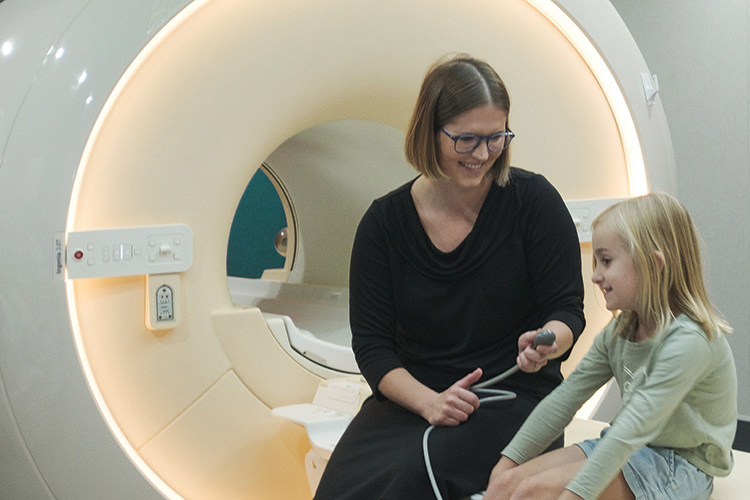
The MRI procedure will be thoroughly explained to you, and your safety questionnaire reviewed and discussed before you enter the MRI scan room. You will usually be asked to change into a gown. You will be asked to lie on the scan table and given a buzzer to hold. When you squeeze it, an alarm sounds in the control room and you will be able to talk to the MRI technologist.
You will not feel the magnetic field or radio waves, so the procedure itself is painless. However, there may be a lot of loud thumping or tapping noises during the scan so we can offer headphones, music or earplugs to help block the sound.
If you are claustrophobic and find you are unable to proceed with the scan you can alert the MRI technologist straight away. The radiology facility has special procedures for people with claustrophobia and will advise you of what to do if this applies to you. MRI images are very susceptible to movement, so it is important that you are comfortable enough to lie still and hold your position during the scan.
Prior to your scan and to ensure optimal imaging, you will be asked to change into a gown. A change cubicle will be provided to ensure your privacy. As the machine being used for this scan contains a magnet, you will be asked to remove all metal items, such as hair clips, jewellery, hearing aids, dentures. A lockable cupboard will be provided for you to securely store all your belongings for the duration of your scan. After your scan, you will be provided with a change cubicle to ensure your privacy. Please ensure you have all your belongings with you prior to leaving the department. If you accidentally leave anything behind, please contact our staff to advise and we will endeavour to locate your belongings and return them to you.
The scan can take between 15 minutes to over 60 minutes to complete. This depends on the part of the body being imaged and what type of MRI is required to show the information. Before the scan begins, the radiographer will tell you how long the scan takes, so you know what to expect.
Our friendly reception staff will advise you on the cost of your procedure at the time of your booking.

